I use a combination of internal and external laboratories to help me conduct my analyses. When I started at Weber in 2016 we were about to move into a new science building that included funding for new lab spaces. I helped make purchases and set up our rock crushing and separation facilities to allow us to separate for zircons and apatites. In 2023 I wrote a proposal with colleagues in Physics and Chemistry to procure a new cathodoluminescence detector for our scanning electron microscope to increase our analytical capabilities, specifically so we could image complex zircons to help us target for LA-ICP-MS analysis at the University of Arizona Laserchron Center.
I most commonly use U-Pb dating of detrital zircons to better understand basin evolution. Along with U-Pb, I also employ Lu/Hf isotopic analysis and trace elements of zircons to fingerprint sources and determine petrogenetics of whichever system I am studying. When zircons are not available I will employ U-Pb on apatite, when available. We are now set up for all separations and imaging at WSU with analyses being completed by my students and me at the University of Arizona Laserchron Center for LA-ICP-MS and Boise State University for ID-TIMS.

A zircon mount.

Dr. Rabosky (Physics) and I with a rock sample and zircon mount infront of the Sem.
If I want to dig into the time-temperature history of a system in more detail I will move into lower temperature systems such as apatite or zircon U-Th/He and fission track. Work in these systems has been completed by myself or colleagues at other institutions including the University of Arizona, Indiana and Illinois.
Zircon Separation and Imaging Facilities
Rock Crushing and Mineral Separations Lab
Our rock crushing and separations labs include sledge and rock hammers for breaking samples, a chipmunk jaw crusher, a disk mill, sieves, Frantz magnetic separators, a gemini water table and hood and equipment for heavy liquid separation. I have trained undergraduates and graduate students from nearby universities in zircon and apatite separation techniques.
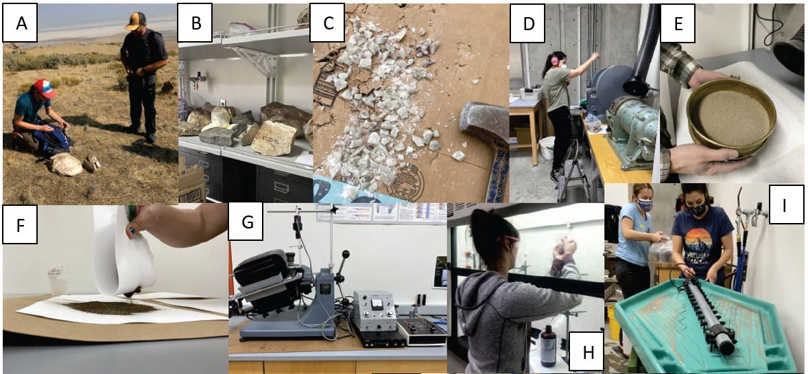
Steps in the process from collecting a sample (A), crushing the sample (B-D), sieving (E), completing magnetic (F and G) and density separations (H and I).
Scanning Electron Microscope with Cathodoluminescence Detector
We have a Thermo FEI Quanta 250 scanning electron microscope with a new The Gatan Monarch Pro CL System. This is a multi-user lab facility, but I primarily use the SEM for imaging thin sections, rock fragments and zircon mounts. We have a Anatech HUMMER 6.2 performs sputter coating system we use to carbon coat mounts for better imaging in the SEM.

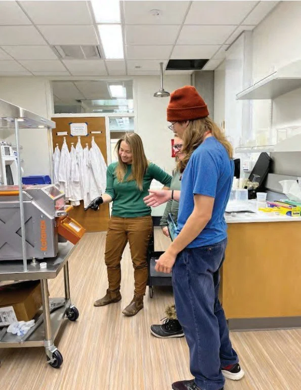
new CL detecror in our SEM lab with the PI’s who wrote the grant.
We requested and received funding for the purchase of a multispectral, high-resolution cathodoluminescence (CL) system that supports ongoing projects and will enhance the next generation of research studies at Weber State. The Gatan Monarch Pro CL System is now installed on our FEI Quanta 250 SEM in the College of Science Microscopy Center (COSMiC) in Tracy Hall Science Center. Installation occurred between May and June of 2024 and faculty and students were trained on the instrument by Gatan technicians in August and September. The CL system is already being utilized to promote student training in instrumentation, data science, and image processing via undergraduate research experiences and course-based projects. The system has significantly enhanced research infrastructure and capabilities, supporting interdisciplinary collaborations among faculty and students in multiple science departments at WSU and with outside institutions and government agencies.
Scanning electron microscopy cathodoluminescence (SEM-CL) has become a key micro-analytical technique to image features within a variety of materials, which are not observable by other methods. The interaction of an electron beam with a sample generates multiple signals for micro-analysis, including secondary electrons related to surface morphology, backscattered electrons (BSE) related to mean atomic number, characteristic X-rays related to elemental composition, and cathodoluminescence (CL) related to defects and trace element chemistry. The use of an electron beam in a SEM allows finer spatial resolution compared to standard petrographic microscopy. SEM-CL systems provide the wavelength resolution of CL spectra necessary to characterize specific trace elements and lattice defects, and can be combined with beam scanning to generate hyperspectral CL maps. Such CL maps can then be compared to SEM BSE and X-ray maps, and integrated with other techniques, including laser-ablation inductively-coupled-mass-spectrometry (LA-ICPMS) analysis of isotopes and trace elements, electron-microprobe mapping, and micro-Raman spectroscopic analysis, to quantify spatial distributions of trace elements and defects within complex materials.
Active projects at WSU that are utilizing integrated CL imaging include: (1) geochronologic and geochemical analysis of minerals to test models of crustal evolution of Paleoproterozoic basement rocks with inherited Archean components western North America (Figure 3), which record a key time in Earth history marked by onset of supercontinent cycles along with the characterization of source-to-sink patterns in distal sediments sourced from continental collisions, which will increase understanding of sediment production and distributions system, and imaging igneous zircons associated with the Yellowstone Hotspot Track to understand chemical evolution of continental hotspots. (2) The CL system is being used for a collaborative research project between the department of Earth & Environmental Sciences (EES), Physics, and Chemistry in which Chemistry is creating synthetic zircon crystals to be irradiated externally by an alpha source in order to damage the surface of the crystals in a similar manner to geologic degradation. The damage is being analyzed via the CL in order to calibrate surface damage to the amount of radiation the grains are exposed to.
Petrographic and Optical Microscopy
Geochemistry Facilities
X-Ray Florescence Spectroscopy
We have a ball mill for crushing rock samples to a powder and all equipment to make fused beads for major element analysis and pressed pellets for trace element analysis. Following processing samples can be loaded into the Rigaku SuperMini200 X-Ray Fluorescence spectrometer.

Student weighing out rock powder and a flux to make a glass bead for geochemical analysis.
Watching the beads being made in the fusion furnace.